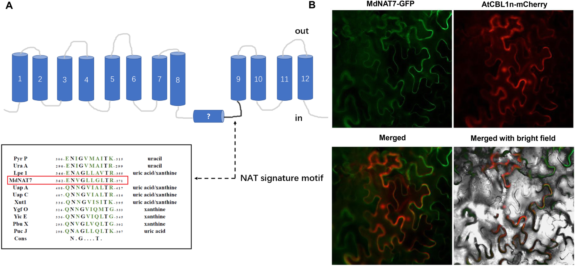
Fig. 1

Analysis and localization of MdNAT7. a. Model of the transmembrane structure of NAT proteins and the NAT signature motif. Pyr P, Bacillus subtilis Pyr P (P31466), Ura A, Escherichia coli homologue Ura A (P0AGM7); Lpe 1, maize Leaf Permease 1 (NP_001150400.1); MdNAT7, M. domestica NAT7 (MDP0000304285); Uap A, Aspergillus nidulans Uap A (Q07307); Uap C, A. nidulans Uap C (P48777); Xut1, Candida albicans Xut1 (AAX22221.1); Ygf O, E. coli homologue YgfO (P67444); Yic E, E. coli homologue Yic E (P0AGM9); Pbu X, B. subtilis Pbu X (P42086); Puc J, B. subtilis Pbu J (O32139); Cons, Consensus refers to the nucleobase-ascorbate transporter motif. b. Localization of MdNAT7. The fusion protein of MdNAT7–GFP was transiently expressed in tobacco leaves and observed with confocal microscopy. MdNAT7–GFP fluorescence. The plasma membrane protein localization marker AtCBL1n-mCherry. Merged images. Merged with bright field. Scale bar = 50 μm